
2D Echo Cost in Bengaluru - Heart Scan Price and What It Shows
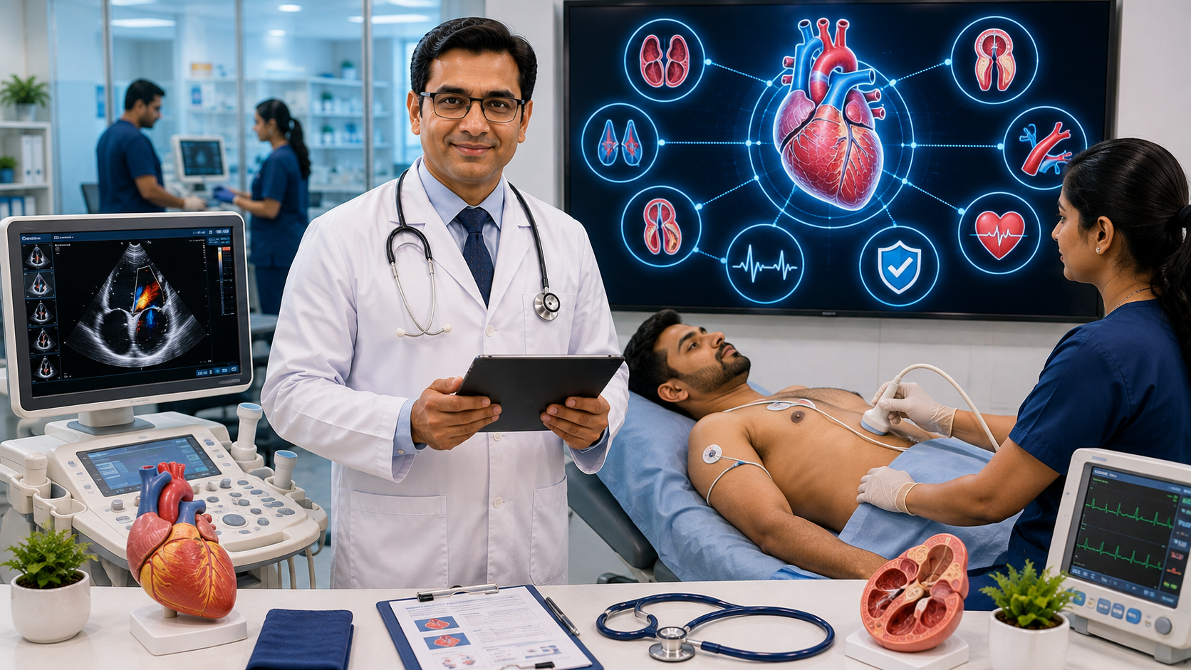
2D Echo Cost in Bengaluru and What It Shows
Looking for the 2D echo cost in Bengaluru and wondering whether you need this heart scan? A 2D echocardiogram (2D Echo) is a safe, non-invasive ultrasound scan that creates real-time images of your heart. It helps doctors evaluate the heart's chambers, valves, pumping function, blood flow, and surrounding structures. The 2D echo cost in Bengaluru typically varies depending on the diagnostic centre, equipment, cardiologist expertise, and whether additional cardiac investigations are recommended. It is commonly advised for people with chest pain, breathlessness, heart murmurs, high blood pressure, suspected heart disease, or those requiring follow-up after a cardiac diagnosis.
Heart disease remains one of the leading causes of illness worldwide. Fortunately, modern imaging techniques allow doctors to detect many heart problems before they become life-threatening. Among these investigations, the 2D Echo is one of the most valuable because it provides detailed information about the heart's structure and function without exposing patients to radiation. Therefore, understanding its purpose, cost, and benefits can help you make informed healthcare decisions.
This Blog Includes:
 What Is a 2D Echo?
A 2D Echocardiogram, commonly called a 2D Echo, is an ultrasound examination of the heart.
Instead of using X-rays, it uses high-frequency sound waves to produce moving images of the heart in real time.
As a result, cardiologists can observe how your heart beats, how blood flows through the chambers, and whether the valves are functioning properly.
Moreover, the procedure is painless, non-invasive, and does not involve radiation exposure.
How Does a 2D Echo Work?
During the scan, a trained cardiac sonographer or cardiologist places a small handheld device called a transducer on the chest.
The transducer emits ultrasound waves that bounce off the heart structures.
A computer then converts these reflected waves into live images displayed on a monitor.
Consequently, doctors can assess both the anatomy and function of the heart during the examination.
Why Is a 2D Echo Important?
A 2D Echo provides valuable information that cannot be obtained through a routine physical examination alone.
It helps doctors:
What Is a 2D Echo?
A 2D Echocardiogram, commonly called a 2D Echo, is an ultrasound examination of the heart.
Instead of using X-rays, it uses high-frequency sound waves to produce moving images of the heart in real time.
As a result, cardiologists can observe how your heart beats, how blood flows through the chambers, and whether the valves are functioning properly.
Moreover, the procedure is painless, non-invasive, and does not involve radiation exposure.
How Does a 2D Echo Work?
During the scan, a trained cardiac sonographer or cardiologist places a small handheld device called a transducer on the chest.
The transducer emits ultrasound waves that bounce off the heart structures.
A computer then converts these reflected waves into live images displayed on a monitor.
Consequently, doctors can assess both the anatomy and function of the heart during the examination.
Why Is a 2D Echo Important?
A 2D Echo provides valuable information that cannot be obtained through a routine physical examination alone.
It helps doctors:
- Article Title: 2D Echo Cost in Bengaluru – Heart Scan Price and What It Shows
- Publisher: Prima Diagnostics
- Type: Test Cost Guide Healthcare Blog
- Topics Covered: 2D Echocardiogram, Heart Scan, Heart Valve Assessment, Cardiac Imaging, Heart Pumping Function, Ejection Fraction, ECG vs 2D Echo, TMT vs 2D Echo, Heart Disease Evaluation, Preventive Cardiology, and Patient Education.
- Services Covered: 2D Echocardiography, ECG, Treadmill Test (TMT), Cardiac Risk Assessment, Preventive Heart Health Check-ups, Cardiology Consultation Support, Digital Reports, and Diagnostic Imaging Services.
- Tests Included: 2D Echocardiogram (2D Echo), Electrocardiogram (ECG), Treadmill Test (TMT), Lipid Profile, High-Sensitivity C-Reactive Protein (hs-CRP), Blood Pressure Assessment, Blood Sugar Testing, Cardiac Risk Markers, and Additional Cardiac Investigations as Recommended by Healthcare Providers.
- Educational Guidance: Explains what a 2D Echo is, how it works, what it reveals about heart structure and function, differences between 2D Echo, ECG, and TMT, common indications for the test, preparation, booking process, and the importance of early heart disease detection.
- Preparation Guidance: A routine transthoracic 2D Echo generally does not require special preparation or fasting. Follow any specific instructions given by your healthcare provider. Wear comfortable clothing, carry previous cardiac reports if available, and inform the healthcare team about current medications and any existing heart conditions.
- Service Focus: Cardiac Imaging, Heart Disease Diagnosis, Preventive Cardiology, Heart Health Screening, Diagnostic Imaging, Digital Reporting, and Patient Education.
- FAQ Section: Included.
What Is a 2D Echo?
A 2D Echocardiogram, commonly called a 2D Echo, is an ultrasound examination of the heart.
Instead of using X-rays, it uses high-frequency sound waves to produce moving images of the heart in real time.
As a result, cardiologists can observe how your heart beats, how blood flows through the chambers, and whether the valves are functioning properly.
Moreover, the procedure is painless, non-invasive, and does not involve radiation exposure.
How Does a 2D Echo Work?
During the scan, a trained cardiac sonographer or cardiologist places a small handheld device called a transducer on the chest.
The transducer emits ultrasound waves that bounce off the heart structures.
A computer then converts these reflected waves into live images displayed on a monitor.
Consequently, doctors can assess both the anatomy and function of the heart during the examination.
Why Is a 2D Echo Important?
A 2D Echo provides valuable information that cannot be obtained through a routine physical examination alone.
It helps doctors:
- Assess heart pumping efficiency
- Evaluate heart valve function
- Detect structural abnormalities
- Identify congenital heart defects
- Assess fluid around the heart
- Evaluate heart enlargement
- Monitor known heart conditions
- Diagnostic centre facilities
- Cardiologist expertise
- Ultrasound technology
- Location within Bengaluru
- Whether additional cardiac investigations are advised
- Health packages that include cardiac screening
- Heart valve disease
- Heart failure
- Cardiomyopathy
- Congenital heart disease
- Pericardial effusion
- Enlarged heart chambers
- Pulmonary hypertension (when assessed with Doppler findings)
- Previous heart attack-related heart muscle damage
- Heart structure
- Heart valves
- Pumping function
- Blood flow
- Chamber size
- Structural heart disease
- Valve disorders
- Heart failure evaluation
- Congenital abnormalities
- Electrical activity of the heart
- Heart rhythm
- Conduction abnormalities
- Arrhythmias
- Acute heart attack evaluation
- Rhythm monitoring
- Heart performance during exercise
- Exercise-induced reduced blood supply to the heart
- Suspected coronary artery disease
- Exercise-related chest pain
- Fitness assessment in selected patients
- Adults over 40 years
- People with diabetes
- Individuals with high blood pressure
- Smokers
- People with high cholesterol
- Those with obesity
- Individuals with a family history of heart disease
- Patients experiencing unexplained fatigue or breathlessness
- Schedule an appointment online
- Call the diagnostic centre
- Visit the centre directly
- Book through your physician's referral